
3D Imaging refers to the technology that creates three-dimensional visual representations of the body’s internal structures. Unlike traditional 2D imaging, which provides flat images, 3D Imaging offers depth and clarity, giving doctors a more accurate view of organs, bones, tissues, and other anatomical features. This advanced visualization allows medical professionals to diagnose, plan treatments, and perform procedures with higher precision.
3D Imaging plays a crucial role in modern medical diagnostics. It enhances the ability to detect diseases early, understand complex anatomies, and monitor conditions over time. This technology improves outcomes by reducing errors and unnecessary invasive procedures. From cancer detection to dental restorations like fitting a tooth cap, 3D Imaging has become indispensable.
How Does 3D Imaging Improve Diagnostic Accuracy?
Traditional diagnostic tools often miss subtle abnormalities because they present only flat, two-dimensional images. In contrast, 3D Imaging provides a volumetric view, enabling doctors to examine an organ or tissue from multiple angles. This multi-angle perspective reveals details that would otherwise remain hidden.
For example, in oncology, 3D scans allow oncologists to pinpoint tumor size, shape, and exact location, facilitating targeted treatment plans. In cardiology, 3D Imaging helps visualize heart valves and vessels, enabling precise assessment of defects or blockages. This improved accuracy reduces misdiagnosis and helps tailor treatment strategies.
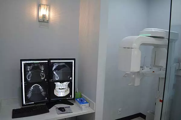
In What Ways Has 3D Imaging Impacted Dentistry, Especially for Tooth Cap Procedures?
Dentistry benefits significantly from 3D Imaging, particularly in restorative procedures such as placing a tooth cap. A tooth cap, or dental crown, protects a damaged tooth and restores its shape and function. Accurate fitting of a tooth cap is vital for comfort and long-term success.
3D Imaging enables dentists to create detailed digital models of a patient’s teeth and jaw. Using these models, they can design perfectly fitting tooth cap that align seamlessly with natural teeth. This technology eliminates the guesswork common in traditional molds and impressions, resulting in faster procedures, fewer adjustments, and enhanced patient satisfaction.
Moreover, 3D Imaging in dentistry supports guided implant surgeries and helps detect cavities or structural issues early, making treatments more effective and minimally invasive.
How Has 3D Imaging Enhanced Surgical Planning and Execution?
Surgical planning has been transformed by the introduction of 3D Imaging. Surgeons can now visualize complex anatomy before making any incisions. By examining a 3D model of the affected area, they understand the exact location and relation of critical structures such as nerves, blood vessels, and organs.
This advance is particularly crucial in delicate surgeries such as brain, spine, or heart operations. It reduces the risk of complications and shortens surgery time. Additionally, 3D Imaging can guide surgeons in real-time, improving precision and patient safety.
What Are the Different Types of 3D Imaging Technologies Used in Medical Diagnostics?
Several types of 3D Imaging technologies have revolutionized medical diagnostics, each suited to specific applications:
- CT Scans (Computed Tomography): CT uses X-rays to generate detailed cross-sectional images of the body. These images combine to form a comprehensive 3D view, commonly used in trauma, oncology, and cardiology.
- MRI (Magnetic Resonance Imaging): MRI employs magnetic fields and radio waves to create 3D images of soft tissues. It excels in imaging the brain, muscles, joints, and heart.
- Ultrasound with 3D Capability: Traditional ultrasound is 2D, but 3D ultrasound offers volumetric imaging, particularly useful in obstetrics and gynecology.
- Cone Beam CT (CBCT): CBCT is widely used in dentistry to provide 3D images of teeth, bone, and soft tissues, aiding procedures like tooth cap placement and implant planning.
Each technology has unique strengths, and medical professionals choose based on the diagnostic need.
How Does 3D Imaging Reduce Patient Risk and Improve Outcomes?
3D Imaging minimizes patient risk by decreasing the need for exploratory surgeries and unnecessary biopsies. With clearer visualization, doctors can avoid damaging vital structures during interventions. Additionally, 3D Imaging enables less invasive procedures, which lead to quicker recoveries and less post-operative pain.
Patients also benefit from personalized treatments based on precise anatomical data, which improves success rates. For instance, custom-designed implants or prosthetics, such as tooth caps created with 3D data, fit better and last longer.
How Has 3D Imaging Made Medical Diagnostics More Accessible?
Technological advancements have reduced the size and cost of 3D Imaging equipment, making it accessible in smaller clinics and remote areas. Portable 3D ultrasound devices, for example, enable point-of-care diagnostics in emergency rooms and field hospitals.
Furthermore, 3D Imaging data can be shared digitally with specialists worldwide, enabling collaborative diagnosis and consultation. This global access improves diagnostic quality and speeds up treatment decisions.
What Role Does 3D Imaging Play in Monitoring Chronic Conditions?
Chronic diseases require continuous monitoring to adjust treatments effectively. 3D Imaging allows doctors to track disease progression visually and quantitatively. For example, in arthritis patients, 3D scans can measure joint degradation over time.
In dental care, regular 3D scans help dentists monitor the integrity of a tooth cap and surrounding teeth, ensuring early detection of issues like decay or misalignment.
How Is 3D Imaging Shaping the Future of Personalized Medicine?
The precision of 3D Imaging supports the development of personalized medicine. By combining 3D anatomical data with genetic and biochemical information, healthcare providers tailor treatments specifically to the individual’s unique profile.
This approach optimizes drug dosages, surgical techniques, and rehabilitation plans, maximizing efficacy and minimizing side effects. In dentistry, personalized tooth caps and implants created from 3D scans exemplify this trend, improving function and aesthetics.
What Challenges Remain in the Adoption of 3D Imaging?
Despite its benefits, 3D Imaging faces challenges, including high costs, the need for specialized training, and large data storage requirements. Some patients may also face exposure to radiation depending on the technology used, though newer methods continually reduce this risk.
Efforts to make 3D Imaging more affordable and user-friendly continue, and innovations in AI-assisted image analysis are helping to streamline workflows and improve diagnostic speed.
In conclusion, 3D Imaging has fundamentally changed medical diagnostics by providing detailed, accurate, and actionable images. From improving surgical outcomes to revolutionizing dental procedures like tooth cap placement, it enhances patient care across disciplines. As technology advances, the role of 3D Imaging in medicine will only grow, offering ever more precise, personalized, and effective diagnostics.